

Which Of The Following Represents An External Control Of The Cell Cycle?

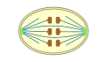
Mitosis in an brute cell (phases ordered counter-clockwise).


Onion (Allium) cells in different phases of the cell cycle enlarged 800 diameters.
a. non-dividing cells
b. nuclei preparing for division (spireme-stage)
c. dividing cells showing mitotic figures
e. pair of daughter-cells shortly after segmentation
In cell biology, mitosis () is a part of the cell cycle in which replicated chromosomes are separated into two new nuclei. Cell division past mitosis gives rise to genetically identical cells in which the total number of chromosomes is maintained.[1] Therefore, mitosis is also known as equational division.[ii] [3] In general, mitosis is preceded by S phase of interphase (during which DNA replication occurs) and is often followed by telophase and cytokinesis; which divides the cytoplasm, organelles and cell membrane of one cell into two new cells containing roughly equal shares of these cellular components.[four] The unlike stages of mitosis altogether define the mitotic (M) stage of an beast cell cycle—the sectionalisation of the female parent cell into ii girl cells genetically identical to each other.[5]
The procedure of mitosis is divided into stages corresponding to the completion of one set up of activities and the showtime of the side by side. These stages are preprophase (specific to plant cells), prophase, prometaphase, metaphase, anaphase, and telophase. During mitosis, the chromosomes, which have already duplicated, condense and attach to spindle fibers that pull one re-create of each chromosome to opposite sides of the cell.[half dozen] The result is two genetically identical girl nuclei. The residue of the jail cell may then go along to divide past cytokinesis to produce two girl cells.[vii] The unlike phases of mitosis can be visualized in real time, using alive jail cell imaging.[8] Producing three or more than daughter cells instead of the normal two is a mitotic error chosen tripolar mitosis or multipolar mitosis (direct cell triplication / multiplication).[9] Other errors during mitosis tin induce apoptosis (programmed cell expiry) or cause mutations. Certain types of cancer can ascend from such mutations.[10]
Mitosis occurs but in eukaryotic cells. Prokaryotic cells, which lack a nucleus, divide past a unlike process called binary fission[ citation needed ]. Mitosis varies between organisms.[xi] For example, animate being cells undergo an "open" mitosis, where the nuclear envelope breaks down before the chromosomes dissever, whereas fungi undergo a "closed" mitosis, where chromosomes divide within an intact jail cell nucleus.[12] Most animal cells undergo a shape change, known every bit mitotic cell rounding, to adopt a near spherical morphology at the start of mitosis. Most human cells are produced by mitotic prison cell partition. Of import exceptions include the gametes – sperm and egg cells – which are produced past meiosis.
Discovery [edit]
Numerous descriptions of cell division were made during 18th and 19th centuries, with various degrees of accuracy.[13] In 1835, the German botanist Hugo von Mohl, described cell division in the green algae Cladophora glomerata, stating that multiplication of cells occurs through jail cell partitioning.[14] [15] [16] In 1838, Matthias Jakob Schleiden affirmed that "formation of new cells in their interior was a full general rule for prison cell multiplication in plants", a view later rejected in favour of Mohl'southward model, due to contributions of Robert Remak and others.[17]
In creature cells, cell division with mitosis was discovered in frog, rabbit, and cat cornea cells in 1873 and described for the first time by the Polish histologist Wacław Mayzel in 1875.[18] [19]
Bütschli, Schneider and Fol might have besides claimed the discovery of the process presently known as "mitosis".[13] In 1873, the High german zoologist Otto Bütschli published data from observations on nematodes. A few years later, he discovered and described mitosis based on those observations.[20] [21] [22]
The term "mitosis", coined by Walther Flemming in 1882,[23] is derived from the Greek discussion μίτος (mitos, "warp thread").[24] [25] There are some alternative names for the process,[26] e.one thousand., "karyokinesis" (nuclear segmentation), a term introduced by Schleicher in 1878,[27] [28] or "equational sectionalization", proposed by August Weismann in 1887.[29] Nonetheless, the term "mitosis" is also used in a broad sense by some authors to refer to karyokinesis and cytokinesis together.[30] Shortly, "equational division" is more commonly used to refer to meiosis II, the part of meiosis most similar mitosis.[31]
Phases [edit]
Overview [edit]
The chief outcome of mitosis and cytokinesis is the transfer of a parent cell's genome into two daughter cells. The genome is equanimous of a number of chromosomes—complexes of tightly coiled Deoxyribonucleic acid that contain genetic information vital for proper jail cell function.[32] Because each resultant daughter cell should exist genetically identical to the parent cell, the parent cell must brand a copy of each chromosome earlier mitosis. This occurs during the S phase of interphase.[33] Chromosome duplication results in two identical sister chromatids bound together by cohesin proteins at the centromere.
When mitosis begins, the chromosomes condense and become visible. In some eukaryotes, for case animals, the nuclear envelope, which segregates the Dna from the cytoplasm, disintegrates into small vesicles. The nucleolus, which makes ribosomes in the jail cell, also disappears. Microtubules project from opposite ends of the cell, attach to the centromeres, and align the chromosomes centrally within the cell. The microtubules then contract to pull the sister chromatids of each chromosome apart.[34] Sister chromatids at this point are called girl chromosomes. As the cell elongates, corresponding daughter chromosomes are pulled toward opposite ends of the cell and condense maximally in late anaphase. A new nuclear envelope forms around the separated daughter chromosomes, which decondense to form interphase nuclei.
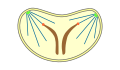
During mitotic progression, typically afterward the anaphase onset, the jail cell may undergo cytokinesis. In animal cells, a jail cell membrane pinches inward between the 2 developing nuclei to produce two new cells. In constitute cells, a cell plate forms between the two nuclei. Cytokinesis does not e'er occur; coenocytic (a type of multinucleate condition) cells undergo mitosis without cytokinesis.
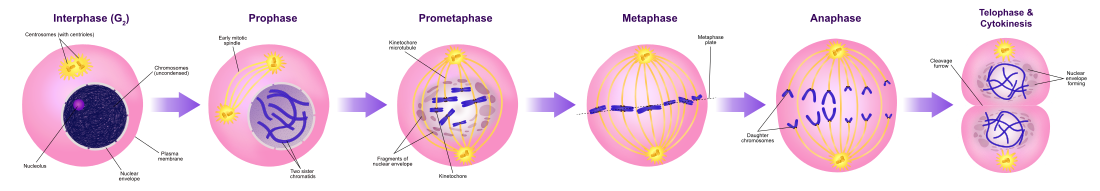
Diagram of the mitotic phases
Interphase [edit]
The mitotic phase is a relatively short menstruation of the jail cell cycle. It alternates with the much longer interphase, where the cell prepares itself for the procedure of cell sectionalization. Interphase is divided into three phases: G1 (first gap), Southward (synthesis), and G2 (second gap). During all iii parts of interphase, the cell grows by producing proteins and cytoplasmic organelles. Yet, chromosomes are replicated only during the S phase. Thus, a jail cell grows (G1), continues to grow as it duplicates its chromosomes (S), grows more and prepares for mitosis (Gtwo), and finally divides (M) before restarting the bike.[33] All these phases in the cell cycle are highly regulated past cyclins, cyclin-dependent kinases, and other cell bicycle proteins. The phases follow one another in strict lodge and there are "checkpoints" that give the cell cues to proceed from 1 phase to some other.[35] Cells may also temporarily or permanently leave the cell cycle and enter M0 phase to stop dividing. This can occur when cells become overcrowded (density-dependent inhibition) or when they differentiate to carry out specific functions for the organism, as is the example for human heart muscle cells and neurons. Some G0 cells accept the ability to re-enter the jail cell wheel.
DNA double-strand breaks can be repaired during interphase past ii principal processes.[36] The first process, not-homologous end joining (NHEJ), tin can join the two broken ends of Dna in the G1, Due south and G2 phases of interphase. The second process, homologous recombinational repair (HRR), is more accurate than NHEJ in repairing double-strand breaks. HRR is active during the S and G2 phases of interphase when Deoxyribonucleic acid replication is either partially accomplished or later on it is completed, since HRR requires ii side by side homologs.
Interphase helps prepare the cell for mitotic segmentation. It dictates whether the mitotic cell division will occur. It carefully stops the cell from proceeding whenever the cell's DNA is damaged or has not completed an of import phase. The interphase is very important every bit it will determine if mitosis completes successfully. Information technology volition reduce the amount of damaged cells produced and the production of malignant cells. A miscalculation by the key Interphase proteins could be crucial every bit the latter could potentially create cancerous cells.[37] Today, more research is existence washed to understand specifically how the phases stated in a higher place occur.
Mitosis [edit]

Stages of early mitosis in a vertebrate jail cell with micrographs of chromatids
Preprophase (plant cells) [edit]
In found cells merely, prophase is preceded past a pre-prophase phase. In highly vacuolated plant cells, the nucleus has to drift into the center of the prison cell before mitosis can begin. This is achieved through the formation of a phragmosome, a transverse sheet of cytoplasm that bisects the cell along the future plane of prison cell sectionalization. In addition to phragmosome formation, preprophase is characterized by the formation of a band of microtubules and actin filaments (called preprophase band) underneath the plasma membrane around the equatorial plane of the futurity mitotic spindle. This band marks the position where the prison cell will eventually split up. The cells of higher plants (such equally the flowering plants) lack centrioles; instead, microtubules form a spindle on the surface of the nucleus and are then organized into a spindle by the chromosomes themselves, after the nuclear envelope breaks downwards.[38] The preprophase ring disappears during nuclear envelope breakdown and spindle formation in prometaphase.[39] : 58–67
Prophase [edit]

Condensing chromosomes. Interphase nucleus (left), condensing chromosomes (eye) and condensed chromosomes (correct).

During prophase, which occurs after G2 interphase, the cell prepares to divide by tightly condensing its chromosomes and initiating mitotic spindle formation. During interphase, the genetic material in the nucleus consists of loosely packed chromatin. At the onset of prophase, chromatin fibers condense into discrete chromosomes that are typically visible at high magnification through a light microscope. In this phase, chromosomes are long, thin, and thread-like. Each chromosome has ii chromatids. The two chromatids are joined at the centromere.
Gene transcription ceases during prophase and does non resume until late anaphase to early on 1000i phase.[twoscore] [41] [42] The nucleolus also disappears during early prophase.[43]
Close to the nucleus of creature cells are structures called centrosomes, consisting of a pair of centrioles surrounded by a loose collection of proteins. The centrosome is the coordinating eye for the jail cell's microtubules. A cell inherits a single centrosome at jail cell partition, which is duplicated by the prison cell before a new round of mitosis begins, giving a pair of centrosomes. The two centrosomes polymerize tubulin to help form a microtubule spindle apparatus. Motor proteins then push the centrosomes forth these microtubules to opposite sides of the cell. Although centrosomes assist organize microtubule associates, they are not essential for the formation of the spindle apparatus, since they are absent from plants,[38] and are not absolutely required for animate being cell mitosis.[44]
Prometaphase [edit]
At the kickoff of prometaphase in animal cells, phosphorylation of nuclear lamins causes the nuclear envelope to disintegrate into small membrane vesicles. As this happens, microtubules invade the nuclear space. This is chosen open up mitosis, and it occurs in some multicellular organisms. Fungi and some protists, such every bit algae or trichomonads, undergo a variation called airtight mitosis where the spindle forms inside the nucleus, or the microtubules penetrate the intact nuclear envelope.[45] [46]
In late prometaphase, kinetochore microtubules brainstorm to search for and attach to chromosomal kinetochores.[47] A kinetochore is a proteinaceous microtubule-bounden structure that forms on the chromosomal centromere during late prophase.[47] [48] A number of polar microtubules find and interact with corresponding polar microtubules from the opposite centrosome to grade the mitotic spindle.[49] Although the kinetochore construction and function are non fully understood, it is known that information technology contains some form of molecular motor.[fifty] When a microtubule connects with the kinetochore, the motor activates, using energy from ATP to "crawl" up the tube toward the originating centrosome. This motor activity, coupled with polymerisation and depolymerisation of microtubules, provides the pulling strength necessary to later carve up the chromosome's two chromatids.[50]
Metaphase [edit]
A cell in late metaphase. All chromosomes (blue) but one take arrived at the metaphase plate.

After the microtubules have located and attached to the kinetochores in prometaphase, the 2 centrosomes begin pulling the chromosomes towards reverse ends of the cell. The resulting tension causes the chromosomes to align along the metaphase plate or equatorial airplane, an imaginary line that is centrally located between the ii centrosomes (at approximately the midline of the prison cell).[49] To ensure equitable distribution of chromosomes at the terminate of mitosis, the metaphase checkpoint guarantees that kinetochores are properly fastened to the mitotic spindle and that the chromosomes are aligned forth the metaphase plate.[51] If the cell successfully passes through the metaphase checkpoint, it proceeds to anaphase.
Anaphase [edit]

During anaphase A, the cohesins that demark sister chromatids together are broken, forming two identical daughter chromosomes.[52] Shortening of the kinetochore microtubules pulls the newly formed girl chromosomes to opposite ends of the prison cell. During anaphase B, polar microtubules push against each other, causing the cell to elongate.[53] In belatedly anaphase, chromosomes also reach their overall maximal condensation level, to help chromosome segregation and the re-germination of the nucleus.[54] In most brute cells, anaphase A precedes anaphase B, but some vertebrate egg cells demonstrate the opposite order of events.[52]
Telophase [edit]

Telophase (from the Greek give-and-take τελος meaning "end") is a reversal of prophase and prometaphase events. At telophase, the polar microtubules continue to lengthen, elongating the jail cell fifty-fifty more. If the nuclear envelope has cleaved down, a new nuclear envelope forms using the membrane vesicles of the parent cell'south old nuclear envelope. The new envelope forms around each set up of separated daughter chromosomes (though the membrane does not enclose the centrosomes) and the nucleolus reappears. Both sets of chromosomes, now surrounded by new nuclear membrane, brainstorm to "relax" or decondense. Mitosis is complete. Each daughter nucleus has an identical fix of chromosomes. Cell sectionalization may or may not occur at this time depending on the organism.
Cytokinesis [edit]

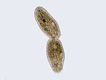
Cilliate undergoing cytokinesis, with the cleavage furrow being clearly visible
Cytokinesis is not a phase of mitosis, but rather a separate process necessary for completing cell division. In animal cells, a cleavage furrow (compression) containing a contractile ring, develops where the metaphase plate used to be, pinching off the separated nuclei.[55] In both animal and institute cells, jail cell division is also driven by vesicles derived from the Golgi apparatus, which move along microtubules to the eye of the cell.[56] In plants, this structure coalesces into a prison cell plate at the centre of the phragmoplast and develops into a cell wall, separating the two nuclei. The phragmoplast is a microtubule structure typical for higher plants, whereas some dark-green algae employ a phycoplast microtubule array during cytokinesis.[39] : 64–seven, 328–9 Each girl prison cell has a complete copy of the genome of its parent cell. The end of cytokinesis marks the stop of the One thousand-phase.
There are many cells where mitosis and cytokinesis occur separately, forming single cells with multiple nuclei. The almost notable occurrence of this is among the fungi, slime molds, and coenocytic algae, only the phenomenon is found in various other organisms. Even in animals, cytokinesis and mitosis may occur independently, for instance during certain stages of fruit fly embryonic evolution.[57]
Function [edit]
Mitosis's "office" or significance relies on the maintenance of the chromosomal set; each formed cell receives chromosomes that are alike in limerick and equal in number to the chromosomes of the parent cell.
Mitosis occurs in the following circumstances:
- Development and growth: The number of cells inside an organism increases by mitosis. This is the footing of the development of a multicellular body from a single prison cell, i.e., zygote and also the ground of the growth of a multicellular body.
- Jail cell replacement: In some parts of the body, due east.yard. pare and digestive tract, cells are constantly sloughed off and replaced by new ones. New cells are formed past mitosis and then are exact copies of the cells being replaced. In like manner, red blood cells have a short lifespan (just about iv months) and new RBCs are formed by mitosis[ citation needed ].
- Regeneration: Some organisms tin can regenerate body parts. The production of new cells in such instances is achieved past mitosis. For example, starfish regenerate lost arms through mitosis.
- Asexual reproduction: Some organisms produce genetically similar offspring through asexual reproduction. For example, the hydra reproduces asexually past budding. The cells at the surface of hydra undergo mitosis and class a mass called a bud. Mitosis continues in the cells of the bud and this grows into a new private. The aforementioned division happens during asexual reproduction or vegetative propagation in plants.
Variations [edit]
Forms of mitosis [edit]
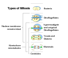
The mitosis process in the cells of eukaryotic organisms follows a similar pattern, but with variations in three main details. "Closed" and "open" mitosis can exist distinguished on the basis of nuclear envelope remaining intact or breaking downwardly. An intermediate form with partial degradation of the nuclear envelope is chosen "semiopen" mitosis. With respect to the symmetry of the spindle apparatus during metaphase, an approximately axially symmetric (centered) shape is called "orthomitosis", distinguished from the eccentric spindles of "pleuromitosis", in which mitotic apparatus has bilateral symmetry. Finally, a 3rd criterion is the location of the central spindle in instance of closed pleuromitosis: "extranuclear" (spindle located in the cytoplasm) or "intranuclear" (in the nucleus).[11]
-
airtight
intranuclear
pleuromitosis -

airtight
extranuclear
pleuromitosis -
airtight
orthomitosis -

semiopen
pleuromitosis -

semiopen
orthomitosis -

open
orthomitosis
Nuclear sectionalisation takes identify but in cells of organisms of the eukaryotic domain, every bit bacteria and archaea have no nucleus. Bacteria and archaea undergo a unlike type of division.[ citation needed ]Inside each of the eukaryotic supergroups, mitosis of the open up course can be found, every bit well as airtight mitosis, except for Excavata, which show exclusively closed mitosis.[58] Following, the occurrence of the forms of mitosis in eukaryotes:[eleven] [59]
- Closed intranuclear pleuromitosis is typical of Foraminifera, some Prasinomonadida, some Kinetoplastida, the Oxymonadida, the Haplosporidia, many fungi (chytrids, oomycetes, zygomycetes, ascomycetes), and some Radiolaria (Spumellaria and Acantharia); it seems to be the virtually primitive blazon.
- Closed extranuclear pleuromitosis occurs in Trichomonadida and Dinoflagellata.
- Closed orthomitosis is found among diatoms, ciliates, some Microsporidia, unicellular yeasts and some multicellular fungi.
- Semiopen pleuromitosis is typical of most Apicomplexa.
- Semiopen orthomitosis occurs with dissimilar variants in some amoebae (Lobosa) and some greenish flagellates (e.g., Raphidophyta or Volvox).
- Open up orthomitosis is typical in mammals and other Metazoa, and in land plants; but it also occurs in some protists.
Errors and other variations [edit]
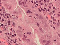
An abnormal (tripolar) mitosis (12 o'clock position) in a precancerous lesion of the stomach (H&E stain)
Errors can occur during mitosis, specially during early embryonic development in humans.[60] During each step of mitosis, there are normally checkpoints too that command the normal outcome of mitosis.[61] But, occasionally to almost rarely, mistakes will happen. Mitotic errors tin can create aneuploid cells that have too few or also many of ane or more chromosomes, a status associated with cancer.[62] [63] Early on human embryos, cancer cells, infected or intoxicated cells can also suffer from pathological sectionalisation into 3 or more daughter cells (tripolar or multipolar mitosis), resulting in severe errors in their chromosomal complements.[9]
In nondisjunction, sis chromatids fail to separate during anaphase.[64] One daughter cell receives both sister chromatids from the nondisjoining chromosome and the other cell receives none. As a event, the former cell gets three copies of the chromosome, a condition known every bit trisomy, and the latter will have only one copy, a condition known every bit monosomy. On occasion, when cells experience nondisjunction, they fail to complete cytokinesis and retain both nuclei in 1 cell, resulting in binucleated cells.[65]
Anaphase lag occurs when the movement of one chromatid is impeded during anaphase.[64] This may be acquired past a failure of the mitotic spindle to properly attach to the chromosome. The lagging chromatid is excluded from both nuclei and is lost. Therefore, one of the daughter cells volition be monosomic for that chromosome.
Endoreduplication (or endoreplication) occurs when chromosomes duplicate but the cell does non subsequently carve up. This results in polyploid cells or, if the chromosomes duplicates repeatedly, polytene chromosomes.[64] [66] Endoreduplication is found in many species and appears to be a normal function of development.[66] Endomitosis is a variant of endoreduplication in which cells replicate their chromosomes during S phase and enter, only prematurely stop, mitosis. Instead of being divided into 2 new daughter nuclei, the replicated chromosomes are retained within the original nucleus.[57] [67] The cells and then re-enter Yard1 and S stage and replicate their chromosomes once again.[67] This may occur multiple times, increasing the chromosome number with each round of replication and endomitosis. Platelet-producing megakaryocytes get through endomitosis during cell differentiation.[68] [69]
Amitosis in ciliates and in beast placental tissues results in a random distribution of parental alleles.
Karyokinesis without cytokinesis originates multinucleated cells called coenocytes.
Diagnostic marker [edit]

Mitosis appearances in breast cancer
In histopathology, the mitosis rate (mitotic count or mitotic index) is an important parameter in diverse types of tissue samples, for diagnosis as well as to further specify the aggressiveness of tumors. For instance, there is routinely a quantification of mitotic count in breast cancer nomenclature.[70] The mitoses must exist counted in an area of the highest mitotic activity. Visually identifying these areas, is difficult in tumors with very high mitotic activeness.[71] Also, the detection of atypical forms of mitosis can exist used both as a diagnostic and prognostic marker.[ citation needed ] For example, lag-type mitosis (non-fastened condensed chromatin in the expanse of the mitotic effigy) indicates high take a chance human papillomavirus infection-related Cervical cancer.[ commendation needed ] In order to ameliorate the reproducibilty and accuracy of the mitotic count, automatic image analysis using deep learning-based algorithms take been proposed.[72] However, farther inquiry is needed before those algorithms can be used to routine diagnostics.
-

Normal and singular forms of mitosis in cancer cells. A, normal mitosis; B, chromatin bridge; C, multipolar mitosis; D, ring mitosis; East, dispersed mitosis; F, asymmetrical mitosis; G, lag-blazon mitosis; and H, micronuclei. H&E stain.
[edit]
Cell rounding [edit]

Cell shape changes through mitosis for a typical animal cell cultured on a apartment surface. The jail cell undergoes mitotic cell rounding during spindle assembly so divides via cytokinesis. The actomyosin cortex is depicted in red, DNA/chromosomes royal, microtubules green, and membrane and retraction fibers in black. Rounding also occurs in live tissue, as described in the text.
In animal tissue, most cells round up to a virtually-spherical shape during mitosis.[73] [74] [75] In epithelia and epidermis, an efficient rounding process is correlated with proper mitotic spindle alignment and subsequent correct positioning of daughter cells.[74] [75] [76] [77] Moreover, researchers have institute that if rounding is heavily suppressed it may result in spindle defects, primarily pole splitting and failure to efficiently capture chromosomes.[78] Therefore, mitotic prison cell rounding is thought to play a protective role in ensuring accurate mitosis.[77] [79]
Rounding forces are driven by reorganization of F-actin and myosin (actomyosin) into a contractile homogeneous cell cortex that one) rigidifies the cell periphery[79] [lxxx] [81] and two) facilitates generation of intracellular hydrostatic pressure (up to 10 fold higher than interphase).[82] [83] [84] The generation of intracellular force per unit area is peculiarly critical nether confinement, such equally would be important in a tissue scenario, where outward forces must be produced to round upwards confronting surrounding cells and/or the extracellular matrix. Generation of pressure is dependent on formin-mediated F-actin nucleation[84] and Rho kinase (ROCK)-mediated myosin Two contraction,[lxxx] [82] [84] both of which are governed upstream by signaling pathways RhoA and ECT2[80] [81] through the activity of Cdk1.[84] Due to its importance in mitosis, the molecular components and dynamics of the mitotic actomyosin cortex is an area of active research.
Mitotic recombination [edit]
Mitotic cells irradiated with 10-rays in the G1 phase of the jail cell cycle repair recombinogenic DNA damages primarily by recombination betwixt homologous chromosomes.[85] Mitotic cells irradiated in the G2 phase repair such damages preferentially by sis-chromatid recombination.[85] Mutations in genes encoding enzymes employed in recombination cause cells to have increased sensitivity to being killed past a diverseness of DNA damaging agents.[86] [87] [88] These findings propose that mitotic recombination is an adaptation for repairing DNA damages including those that are potentially lethal.
Evolution [edit]
Some types of cell division in prokaryotes and eukaryotes
There are prokaryotic homologs of all the key molecules of eukaryotic mitosis (east.g., actins, tubulins). Existence a universal eukaryotic property, mitosis probably arose at the base of operations of the eukaryotic tree. As mitosis is less circuitous than meiosis, meiosis may have arisen after mitosis.[89] However, sexual reproduction involving meiosis is also a primitive characteristic of eukaryotes.[90] Thus meiosis and mitosis may both have evolved, in parallel, from ancestral prokaryotic processes.
While in bacterial cell division, after duplication of DNA, two round chromosomes are fastened to a special region of the prison cell membrane, eukaryotic mitosis is ordinarily characterized by the presence of many linear chromosomes, whose kinetochores attaches to the microtubules of the spindle. In relation to the forms of mitosis, closed intranuclear pleuromitosis seems to be the most archaic type, as it is more like to bacterial division.[11]
Gallery [edit]
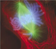
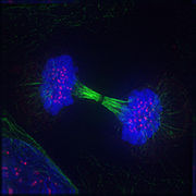
Mitotic cells can be visualized microscopically past staining them with fluorescent antibodies and dyes.
-

Early prophase: Polar microtubules, shown as green strands, take established a matrix around the currently intact nucleus, with the condensing chromosomes in blueish. The cerise nodules are the centromeres.
-

Early prometaphase: The nuclear membrane has simply disassembled, assuasive the microtubules to quickly interact with the kinetochores, which gather on the centromeres of the condensing chromosomes.
-
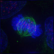
Metaphase: The centrosomes have moved to the poles of the cell and take established the mitotic spindle. The chromosomes have congressed at the metaphase plate.
-

Anaphase: Kinetochore microtubules pull the two sets of chromosomes autonomously, and lengthening polar microtubules button the halves of the dividing cell further apart, while chromosomes are condensed maximally.
-
Telophase: Reversal of prophase and prometaphase events and thus completing the jail cell bicycle.
See too [edit]
- Aneuploidy
- Binary fission
- Chromosome abnormality
- Cytoskeleton
- Meiosis
- Mitogen
- Mitosis Promoting Factor
- Mitotic bookmarking
- Motor protein
References [edit]
- ^ "Cell division and growth". britannica.com. ENCYCLOPÆDIA BRITANNICA. Archived from the original on 2018-ten-28. Retrieved 2018-11-04 .
- ^ "iv.ane: Meiosis". Biology LibreTexts. 2019-10-01. Retrieved 2021-05-29 .
- ^ "Explain why mitosis is called equational and meiosis class 11 biological science CBSE". www.vedantu.com . Retrieved 2021-05-29 .
- ^ Carter JS (2014-01-14). "Mitosis". biology.clc.uc.edu. Archived from the original on 2012-10-27. Retrieved 2019-11-12 .
- ^ "Mitosis - an overview | ScienceDirect Topics". www.sciencedirect.com . Retrieved 2020-11-24 .
- ^ "Cell Sectionalization: Stages of Mitosis | Learn Science at Scitable". www.nature.com. Archived from the original on 2015-11-14. Retrieved 2015-11-16 .
- ^ Maton A, Hopkins JJ, LaHart S, Quon Warner D, Wright K, Jill D (1997). Cells: Building Blocks of Life . New Jersey: Prentice Hall. pp. lxx–4. ISBN978-0-13-423476-2.
- ^ Sandoz PA (December 2019). "Prototype-based analysis of living mammalian cells using label-gratuitous 3D refractive index maps reveals new organelle dynamics and dry out mass flux". PLOS Biological science. 17 (12): e3000553. doi:x.1371/journal.pbio.3000553. PMC6922317. PMID 31856161.
- ^ a b Kalatova B, Jesenska R, Hlinka D, Dudas M (January 2015). "Tripolar mitosis in human cells and embryos: occurrence, pathophysiology and medical implications". Acta Histochemica. 117 (1): 111–25. doi:10.1016/j.acthis.2014.11.009. PMID 25554607.
- ^ Kops GJ, Weaver BA, Cleveland DW (October 2005). "On the road to cancer: aneuploidy and the mitotic checkpoint". Nature Reviews. Cancer. 5 (ten): 773–85. doi:10.1038/nrc1714. PMID 16195750. S2CID 2515388.
- ^ a b c d Raikov IB (1994). "The variety of forms of mitosis in protozoa: A comparative review". European Periodical of Protistology. 30 (three): 253–69. doi:ten.1016/S0932-4739(eleven)80072-6.
- ^ De Souza CP, Osmani SA (September 2007). "Mitosis, non only open or closed". Eukaryotic Cell. half-dozen (9): 1521–7. doi:10.1128/EC.00178-07. PMC2043359. PMID 17660363.
- ^ a b Ross, Anna E. "Human Anatomy & Physiology I: A Chronology of the Clarification of Mitosis". Christian Brothers University. Retrieved 02 May 2018. link Archived 2016-05-12 at the Wayback Auto.
- ^ von Mohl H (1835). Ueber die Vermehrung der Pflanzenzellen durch Theilung. Inaugural-Dissertation (Thesis). Tübingen.
- ^ Karl Mägdefrau (1994), "Mohl, Hugo von", Neue Deutsche Biographie (in German), vol. 17, Berlin: Duncker & Humblot, pp. 690–691 ; (full text online)
- ^ "Notes and memoranda: The late professor von Mohl". Quarterly Journal of Microscopical Science, v. XV, New Series, p. 178-181, 1875. link.
- ^ Weyers, Wolfgang (2002). 150 Years of jail cell division. Dermatopathology: Practical & Conceptual, Vol. 8, No. 2. link Archived 2019-04-02 at the Wayback Auto
- ^ Komender J (2008). "Kilka słów o doktorze Wacławie Mayzlu i jego odkryciu" [On Waclaw Mayzel and his observation of mitotic division] (PDF). Postępy Biologii Komórki (in Shine). 35 (3): 405–407. Archived (PDF) from the original on 2012-10-27.
- ^ Iłowiecki M (1981). Dzieje nauki polskiej. Warszawa: Wydawnictwo Interpress. p. 187. ISBN978-83-223-1876-eight.
- ^ Bütschli, O. (1873). Beiträge zur Kenntnis der freilebenden Nematoden. Nova Acta der Kaiserlich Leopoldinisch-Carolinischen Deutschen Akademie der Naturforscher 36, i-144. link Archived 2018-08-xi at the Wayback Motorcar.
- ^ Bütschli, O. (1876). Studien über die ersten Entwicklungsvorgänge der Eizelle, die Zelleilung und dice Conjugation der Infusorien. Abh.d. Senckenb. Naturf. Ges. Frankfurt a. M. x, 213-452. link Archived 2018-08-09 at the Wayback Machine.
- ^ Fokin SI (2013). "Otto Bütschli (1848–1920) Where nosotros will genuflect?" (PDF). Protistology. viii (1): 22–35. Archived (PDF) from the original on 2014-08-08. Retrieved 2014-08-06 .
- ^ Precipitous LW (1921). Introduction To Cytology. New York: McGraw Hill Volume Company Inc. p. 143.
- ^ "mitosis". Online Etymology Dictionary. Archived from the original on 2017-09-28. Retrieved 2019-xi-12 .
- ^ μίτος . Liddell, Henry George; Scott, Robert; A Greek–English Lexicon at the Perseus Project
- ^ Battaglia Due east (2009). "Caryoneme alternative to chromosome and a new caryological nomenclature" (PDF). Caryologia. 62 (iv): i–fourscore. Archived from the original (PDF) on 2016-03-04.
- ^ Schleicher W (1878). "Dice Knorpelzelltheilung". Arch. Mirkroskop. Anat. 16: 248–300. doi:10.1007/BF02956384. S2CID 163374324. Archived from the original on 2018-08-xi.
- ^ Toepfer G. "Karyokinesis". BioConcepts. Archived from the original on 2018-05-03. Retrieved two May 2018.
- ^ Battaglia Due east (1987). "Embryological questions: 12. Have the Polygonum and Allium types been rightly established?". Ann Bot. Rome. 45: 81–117.
p. 85: Already in 1887, Weismann gave the names Aequationstheilung to the usual prison cell division, and Reduktionstheilungen to the 2 divisions involved in the halving procedure of the number of Kernsegmente
- ^ Mauseth JD (1991). Phytology: an Introduction to Constitute Biology. Philadelphia: Saunders College Publishing. ISBN9780030302220.
p. 102: Cell division is cytokinesis, and nuclear division is karyokinesis. The words "mitosis" and "meiosis" technically refer only to karyokinesis but are often used to describe cytokinesis as well.
- ^ Cooper, Geoffrey M. (2000). "Meiosis and Fertilization". The Cell: A Molecular Approach. 2nd Edition.
- ^ Brown, Terence A. (2002). The Homo Genome. Wiley-Liss.
- ^ a b Blow JJ, Tanaka TU (November 2005). "The chromosome cycle: coordinating replication and segregation. 2nd in the cycles review series". EMBO Reports. 6 (11): 1028–34. doi:10.1038/sj.embor.7400557. PMC1371039. PMID 16264427.
- ^ Zhou J, Yao J, Joshi HC (September 2002). "Zipper and tension in the spindle assembly checkpoint". Journal of Cell Science. 115 (Pt eighteen): 3547–55. doi:10.1242/jcs.00029. PMID 12186941.
- ^ Biology Online (28 Apr 2020). "Mitosis". Biology Online.
- ^ Shibata A (2017). "Regulation of repair pathway choice at two-ended Deoxyribonucleic acid double-strand breaks". Mutat Res. 803–805: 51–55. doi:10.1016/j.mrfmmm.2017.07.011. PMID 28781144.
- ^ Bernat, R. L.; Borisy, Thou. G.; Rothfield, Due north. F.; Earnshaw, W. C. (1990-ten-01). "Injection of anticentromere antibodies in interphase disrupts events required for chromosome movement at mitosis". The Journal of Cell Biology. 111 (four): 1519–1533. doi:x.1083/jcb.111.4.1519. ISSN 0021-9525. PMC2116233. PMID 2211824.
- ^ a b Lloyd C, Chan J (February 2006). "Not and so divided: the common basis of plant and animal cell division". Nature Reviews. Molecular Cell Biology. 7 (2): 147–52. doi:10.1038/nrm1831. PMID 16493420. S2CID 7895964.
- ^ a b Raven PH, Evert RF, Eichhorn SE (2005). Biological science of Plants (7th ed.). New York: W. H. Freeman and Co. ISBN978-0716710073.
- ^ Prasanth KV, Sacco-Bubulya PA, Prasanth SG, Spector DL (March 2003). "Sequential entry of components of the gene expression machinery into daughter nuclei". Molecular Biology of the Cell. 14 (three): 1043–57. doi:x.1091/mbc.E02-10-0669. PMC151578. PMID 12631722.
- ^ Kadauke Due south, Blobel GA (April 2013). "Mitotic bookmarking by transcription factors". Epigenetics & Chromatin. 6 (one): half dozen. doi:10.1186/1756-8935-6-6. PMC3621617. PMID 23547918.
- ^ Prescott DM, Bender MA (March 1962). "Synthesis of RNA and protein during mitosis in mammalian tissue civilization cells". Experimental Cell Inquiry. 26 (2): 260–8. doi:10.1016/0014-4827(62)90176-3. PMID 14488623.
- ^ Olson MO (2011). The Nucleolus. Vol. 15 of Protein Reviews. Berlin: Springer Science & Business organization Media. p. 15. ISBN9781461405146.
- ^ Basto R, Lau J, Vinogradova T, Gardiol A, Woods CG, Khodjakov A, Raff JW (June 2006). "Flies without centrioles". Prison cell. 125 (seven): 1375–86. doi:10.1016/j.cell.2006.05.025. PMID 16814722. S2CID 2080684.
- ^ Heywood P (June 1978). "Ultrastructure of mitosis in the chloromonadophycean alga Vacuolaria virescens". Journal of Cell Science. 31: 37–51. doi:10.1242/jcs.31.1.37. PMID 670329.
- ^ Ribeiro KC, Pereira-Neves A, Benchimol M (June 2002). "The mitotic spindle and associated membranes in the airtight mitosis of trichomonads". Biology of the Prison cell. 94 (3): 157–72. doi:ten.1016/S0248-4900(02)01191-seven. PMID 12206655. S2CID 29081466.
- ^ a b Chan GK, Liu ST, Yen TJ (November 2005). "Kinetochore structure and role". Trends in Cell Biology. 15 (11): 589–98. doi:10.1016/j.tcb.2005.09.010. PMID 16214339.
- ^ Cheeseman IM, Desai A (Jan 2008). "Molecular compages of the kinetochore-microtubule interface". Nature Reviews. Molecular Jail cell Biology. 9 (1): 33–46. doi:10.1038/nrm2310. PMID 18097444. S2CID 34121605.
- ^ a b Winey M, Mamay CL, O'Toole ET, Mastronarde DN, Giddings TH, McDonald KL, McIntosh JR (June 1995). "3-dimensional ultrastructural analysis of the Saccharomyces cerevisiae mitotic spindle". The Journal of Jail cell Biology. 129 (half-dozen): 1601–15. doi:10.1083/jcb.129.six.1601. PMC2291174. PMID 7790357.
- ^ a b Maiato H, DeLuca J, Salmon ED, Earnshaw WC (November 2004). "The dynamic kinetochore-microtubule interface" (PDF). Periodical of Cell Science. 117 (Pt 23): 5461–77. doi:ten.1242/jcs.01536. PMID 15509863. S2CID 13939431. Archived (PDF) from the original on 2017-08-18. Retrieved 2018-04-20 .
- ^ Chan GK, Yen TJ (2003). "The mitotic checkpoint: a signaling pathway that allows a single unattached kinetochore to inhibit mitotic exit". Progress in Cell Cycle Research. five: 431–9. PMID 14593737.
- ^ a b FitzHarris G (March 2012). "Anaphase B precedes anaphase A in the mouse egg" (PDF). Current Biological science. 22 (five): 437–44. doi:10.1016/j.cub.2012.01.041. PMID 22342753. Archived (PDF) from the original on 2018-07-24. Retrieved 2019-09-17 .
- ^ Miller KR, Levine J (2000). "Anaphase". Biology (fifth ed.). Pearson Prentice Hall. pp. 169–lxx. ISBN978-0-13-436265-vi.
- ^ European Molecular Biological science Laboratory (12 June 2007). "Chromosome condensation through mitosis". Science Daily. Archived from the original on 13 June 2007. Retrieved 4 October 2020.
- ^ Glotzer 1000 (March 2005). "The molecular requirements for cytokinesis". Science. 307 (5716): 1735–9. Bibcode:2005Sci...307.1735G. doi:10.1126/science.1096896. PMID 15774750. S2CID 34537906.
- ^ Albertson R, Riggs B, Sullivan W (February 2005). "Membrane traffic: a driving forcefulness in cytokinesis". Trends in Prison cell Biological science. 15 (2): 92–101. doi:10.1016/j.tcb.2004.12.008. PMID 15695096.
- ^ a b Lilly MA, Duronio RJ (April 2005). "New insights into cell cycle command from the Drosophila endocycle". Oncogene. 24 (17): 2765–75. doi:10.1038/sj.onc.1208610. PMID 15838513.
- ^ Boettcher B, Barral Y (2013). "The cell biology of open and closed mitosis". Nucleus. 4 (3): 160–5. doi:10.4161/nucl.24676. PMC3720745. PMID 23644379.
- ^ R. Desalle, B. Schierwater: Key Transitions in Animal Evolution. CRC Printing, 2010, p. 12, link Archived 2019-01-02 at the Wayback Machine.
- ^ Mantikou E, Wong KM, Repping Southward, Mastenbroek South (December 2012). "Molecular origin of mitotic aneuploidies in preimplantation embryos". Biochimica et Biophysica Acta (BBA) - Molecular Basis of Disease. 1822 (12): 1921–thirty. doi:10.1016/j.bbadis.2012.06.013. PMID 22771499.
- ^ Wassmann, Katja; Benezra, Robert (2001-02-01). "Mitotic checkpoints: from yeast to cancer". Current Stance in Genetics & Development. 11 (i): 83–xc. doi:10.1016/S0959-437X(00)00161-1. ISSN 0959-437X. PMID 11163156.
- ^ Draviam VM, Xie S, Sorger PK (Apr 2004). "Chromosome segregation and genomic stability". Current Opinion in Genetics & Development. xiv (two): 120–5. doi:10.1016/j.gde.2004.02.007. PMID 15196457.
- ^ Santaguida S, Amon A (August 2015). "Brusk- and long-term effects of chromosome mis-segregation and aneuploidy". Nature Reviews. Molecular Cell Biological science. xvi (8): 473–85. doi:ten.1038/nrm4025. hdl:1721.one/117201. PMID 26204159. S2CID 205495880.
- ^ a b c Iourov IY, Vorsanova SG, Yurov YB (2006). "Chromosomal Variations in Mammalian Neuronal Cells: Known Facts and Bonny Hypotheses". In Jeon KJ (ed.). International Review Of Cytology: A Survey of Prison cell Biology. Vol. 249. Waltham, MA: Bookish Press. p. 146. ISBN9780080463506.
- ^ Shi Q, King RW (October 2005). "Chromosome nondisjunction yields tetraploid rather than aneuploid cells in human prison cell lines". Nature. 437 (7061): 1038–42. Bibcode:2005Natur.437.1038S. doi:10.1038/nature03958. PMID 16222248. S2CID 1093265.
- ^ a b Edgar BA, Orr-Weaver TL (May 2001). "Endoreplication cell cycles: more than for less". Prison cell. 105 (three): 297–306. doi:x.1016/S0092-8674(01)00334-8. PMID 11348589. S2CID 14368177.
- ^ a b Lee HO, Davidson JM, Duronio RJ (November 2009). "Endoreplication: polyploidy with purpose". Genes & Development. 23 (21): 2461–77. doi:10.1101/gad.1829209. PMC2779750. PMID 19884253.
- ^ Italiano JE, Shivdasani RA (June 2003). "Megakaryocytes and beyond: the birth of platelets". Journal of Thrombosis and Haemostasis. 1 (6): 1174–82. doi:10.1046/j.1538-7836.2003.00290.10. PMID 12871316. S2CID 24325966.
- ^ Vitrat Due north, Cohen-Solal K, Pique C, Le Couedic JP, Norol F, Larsen AK, Katz A, Vainchenker W, Debili N (May 1998). "Endomitosis of human being megakaryocytes are due to abortive mitosis". Blood. 91 (10): 3711–23. doi:x.1182/blood.V91.x.3711. PMID 9573008.
- ^ "Infiltrating Ductal Carcinoma of the Breast (Carcinoma of No Special Blazon)". Stanford University School of Medicine. Archived from the original on 2019-09-11. Retrieved 2019-10-02 .
- ^ Bertram CA, Aubreville 1000, Gurtner C, Bartel A, Corner SM, Dettwiler M, et al. (March 2020). "Computerized Calculation of Mitotic Count Distribution in Canine Cutaneous Mast Cell Tumor Sections: Mitotic Count Is Surface area Dependent" (PDF). Veterinary Pathology. 57 (2): 214–226. doi:10.1177/0300985819890686. PMID 31808382. S2CID 208767801.
- ^ Bertram, Christof A; Aubreville, Marc; Donovan, Taryn A; Bartel, Alexander; Wilm, Frauke; Marzahl, Christian; Assenmacher, Charles-Antoine; Becker, Kathrin; Bennett, Mark; Corner, Sarah; Cossic, Brieuc; Denk, Daniela; Dettwiler, Martina; Gonzalez, Beatriz Garcia; Gurtner, Corinne; Haverkamp, Ann-Kathrin; Heier, Annabelle; Lehmbecker, Annika; Merz, Sophie; Noland, Erika L; Plog, Stephanie; Schmidt, Anja; Sebastian, Franziska; Sledge, Dodd 1000; Smedley, Rebecca C; Tecilla, Marco; Thaiwong, Tuddow; Fuchs-Baumgartinger, Andrea; Meuten, Donald J; Breininger, Katharina; Kiupel, Matti; Maier, Andreas; Klopfleisch, Robert (2021). "Estimator-assisted mitotic count using a deep learning–based algorithm improves interobserver reproducibility and accuracy". Veterinarian Pathology. doi:10.1177/03009858211067478. PMID 34965805. S2CID 245567911.
- ^ Sauer FC (1935). "Mitosis in the neural tube". Journal of Comparative Neurology. 62 (2): 377–405. doi:10.1002/cne.900620207. S2CID 84960254.
- ^ a b Meyer EJ, Ikmi A, Gibson MC (March 2011). "Interkinetic nuclear migration is a broadly conserved feature of cell division in pseudostratified epithelia". Electric current Biology. 21 (6): 485–91. doi:10.1016/j.cub.2011.02.002. PMID 21376598.
- ^ a b Luxenburg C, Pasolli HA, Williams SE, Fuchs East (March 2011). "Developmental roles for Srf, cortical cytoskeleton and cell shape in epidermal spindle orientation". Nature Jail cell Biology. 13 (3): 203–xiv. doi:10.1038/Ncb2163. PMC3278337. PMID 21336301.
- ^ Nakajima Y, Meyer EJ, Kroesen A, McKinney SA, Gibson MC (Baronial 2013). "Epithelial junctions maintain tissue architecture by directing planar spindle orientation". Nature. 500 (7462): 359–62. Bibcode:2013Natur.500..359N. doi:10.1038/nature12335. PMID 23873041. S2CID 4418619.
- ^ a b Cadart C, Zlotek-Zlotkiewicz Eastward, Le Berre M, Piel M, Matthews HK (April 2014). "Exploring the function of cell shape and size during mitosis". Developmental Jail cell. 29 (2): 159–69. doi:10.1016/j.devcel.2014.04.009. PMID 24780736.
- ^ Lancaster OM, Le Berre M, Dimitracopoulos A, Bonazzi D, Zlotek-Zlotkiewicz East, Picone R, Duke T, Piel M, Baum B (May 2013). "Mitotic rounding alters jail cell geometry to ensure efficient bipolar spindle formation". Developmental Cell. 25 (3): 270–83. doi:10.1016/j.devcel.2013.03.014. PMID 23623611.
- ^ a b Lancaster OM, Baum B (October 2014). "Shaping upwardly to divide: coordinating actin and microtubule cytoskeletal remodelling during mitosis". Seminars in Cell & Developmental Biology. 34: 109–xv. doi:ten.1016/j.semcdb.2014.02.015. PMID 24607328.
- ^ a b c Maddox As, Burridge Yard (January 2003). "RhoA is required for cortical retraction and rigidity during mitotic cell rounding". The Journal of Cell Biology. 160 (ii): 255–65. doi:ten.1083/jcb.200207130. PMC2172639. PMID 12538643.
- ^ a b Matthews HK, Delabre U, Rohn JL, Guck J, Kunda P, Baum B (Baronial 2012). "Changes in Ect2 localization couple actomyosin-dependent cell shape changes to mitotic progression". Developmental Jail cell. 23 (two): 371–83. doi:ten.1016/j.devcel.2012.06.003. PMC3763371. PMID 22898780.
- ^ a b Stewart MP, Helenius J, Toyoda Y, Ramanathan SP, Muller DJ, Hyman AA (January 2011). "Hydrostatic pressure and the actomyosin cortex drive mitotic cell rounding". Nature. 469 (7329): 226–30. Bibcode:2011Natur.469..226S. doi:x.1038/nature09642. PMID 21196934. S2CID 4425308.
- ^ Fischer-Friedrich E, Hyman AA, Jülicher F, Müller DJ, Helenius J (August 2014). "Quantification of surface tension and internal pressure level generated by single mitotic cells". Scientific Reports. four (6213): 6213. Bibcode:2014NatSR...4E6213F. doi:10.1038/srep06213. PMC4148660. PMID 25169063.
- ^ a b c d Ramanathan SP, Helenius J, Stewart MP, Cattin CJ, Hyman AA, Muller DJ (February 2015). "Cdk1-dependent mitotic enrichment of cortical myosin Ii promotes cell rounding against confinement". Nature Cell Biology. 17 (2): 148–59. doi:x.1038/ncb3098. PMID 25621953. S2CID 5208968.
- ^ a b Kadyk LC, Hartwell LH (October 1992). "Sis chromatids are preferred over homologs as substrates for recombinational repair in Saccharomyces cerevisiae". Genetics. 132 (2): 387–402. doi:10.1093/genetics/132.2.387. PMC1205144. PMID 1427035.
- ^ Botthof JG, Bielczyk-Maczyńska E, Ferreira L, Cvejic A (May 2017). "rad51 leads to Fanconi anemia-like symptoms in zebrafish". Proceedings of the National Academy of Sciences of the United States of America. 114 (22): E4452–E4461. doi:10.1073/pnas.1620631114. PMC5465903. PMID 28512217.
Here we provide in vivo evidence that the subtract in HSPC numbers in adult fish indeed stems from a combination of decreased proliferation and increased apoptosis during embryonic development. This defect appears to be mediated via p53(10), as our p53/rad51 double mutants did not display whatever observable hematological defects in embryos or adults.
- ^ Stürzbecher HW, Donzelmann B, Henning Westward, Knippschild U, Buchhop S (April 1996). "p53 is linked direct to homologous recombination processes via RAD51/RecA protein interaction". The EMBO Periodical. 15 (8): 1992–2002. doi:10.1002/j.1460-2075.1996.tb00550.ten. PMC450118. PMID 8617246.
- ^ Sonoda Eastward, Sasaki MS, Buerstedde JM, Bezzubova O, Shinohara A, Ogawa H, et al. (Jan 1998). "Rad51-deficient vertebrate cells accumulate chromosomal breaks prior to prison cell death". The EMBO Periodical. 17 (ii): 598–608. doi:10.1093/emboj/17.2.598. PMC1170409. PMID 9430650.
- ^ Wilkins AS, Holliday R (January 2009). "The development of meiosis from mitosis". Genetics. 181 (one): three–12. doi:ten.1534/genetics.108.099762. PMC2621177. PMID 19139151.
- ^ Bernstein, H., Bernstein, C. Evolutionary origin and adaptive function of meiosis. In "Meiosis", Intech Publ (Ballad Bernstein and Harris Bernstein editors), Chapter 3: 41-75 (2013).
Further reading [edit]
- Morgan DL (2007). The cell wheel: principles of command. London: Published by New Science Printing in association with Oxford University Press. ISBN978-0-9539181-2-vi.
- Alberts B, Johnson A, Lewis J, Raff M, Roberts K, Walter P (2002). "Mitosis". Molecular Biology of the Cell (4th ed.). Garland Scientific discipline. Retrieved 2006-01-22 .
- Campbell N, Reece J (December 2001). "The Cell Bike". Biology (6th ed.). San Francisco: Benjamin Cummings/Addison-Wesley. pp. 217–224. ISBN978-0-8053-6624-2.
- Cooper One thousand (2000). "The Events of One thousand Stage". The Jail cell: A Molecular Approach (2nd ed.). Sinaeur Assembly, Inc. Retrieved 2006-01-22 .
- Freeman S (2002). "Prison cell Division". Biology . Upper Saddle River, NJ: Prentice Hall. pp. 155–174. ISBN978-0-13-081923-nine.
- Lodish H, Berk A, Zipursky L, Matsudaira P, Baltimore D, Darnell J (2000). "Overview of the Prison cell Cycle and Its Control". Molecular Cell Biology (quaternary ed.). W. H. Freeman. Retrieved 2006-01-22 .
External links [edit]
| | Wikimedia Eatables has media related to Mitosis. |
- A Flash animation comparison Mitosis and Meiosis
- Khan Academy, lecture
- Studying Mitosis in Cultured Mammalian Cells
- General 1000-12 classroom resources for Mitosis
- The Jail cell-Cycle Ontology
- WormWeb.org: Interactive Visualization of the C. elegans Cell Lineage – Visualize the unabridged cell lineage tree and all of the cell divisions of the nematode C. elegans

Which Of The Following Represents An External Control Of The Cell Cycle?,
Source: https://en.wikipedia.org/wiki/Mitosis
Posted by: lamberttherad.blogspot.com
0 Response to "Which Of The Following Represents An External Control Of The Cell Cycle?"
Post a Comment